Recent research clearly illustrates the potential that artificial intelligence (AI) and deep learning systems offer for improving the detection and diagnosis of cancer, whether from radiological images or tissue analysis. While further clinical validation of such systems is needed before they can be reliably deployed in medical practice, they are expected to benefit patient care by supplementing a physician’s own skill and decision-making ability.
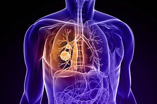 In May, Google’s AI team and researchers from Northwestern University, New York University-Langone Medical Center, and Stanford Medicine published research showing the ability of a cancer-spotting AI system to outperform a group of six radiologists when diagnosing lung cancer from 3-D volumetric CT scans. Radiologists looking for signs of lung cancer normally view hundreds of individual 2-D slices from a single CT scan. In contrast, the AI system examined each lung scan as a single 3-D object, and by comparing it with prior scans from the same patient, could identify subtle lung nodules that are difficult for humans to detect, thus allowing it to generate an overall prediction of malignancy. The researchers showed that when applied to 45,800 chest CT scans, gathered from data sets from the National Institutes of Health and Northwestern, the AI system was able to retrospectively detect cancer with an overall success rate of 94.4%. The AI system outperformed the radiologists, detecting 5% more cancer cases, while reducing false positives by more than 11%. In one case, the AI system diagnosed lung cancer in an asymptomatic patient a year prior to the patient’s cancer diagnosis from a CT scan that had been judged normal. Experts believe the ability to detect cancer as much as 12 months earlier than usual could translate to an increased survival rate as high as 40%.
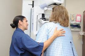 In June, scientists from IBM Research in Israel detailed an AI model capable of identifying patients likely to develop breast cancer within a year, correctly predicting the development of 87% and 77% of malignant and benign tumors, respectively. The system, trained on 9,611 mammograms and patient electronic health records, was also able to identify breast cancer in 48% of people that otherwise would not have been detected, an accuracy comparable to radiologists when a second “reader” is used to confirm the diagnosis. The training set was unique, linking the mammogram images to data from the patient’s electronic health records, including information on thyroid function, reproductive history, white blood cell profiles, metabolic syndrome and other health information. The model mapped connections among the various clinical risk factors for breast cancer to anticipate biopsy malignancy and differentiate normal from abnormal screening scans.
The AI model was also tested on 71 false-negative cases where the patient was later diagnosed with breast cancer within 12 months. In this case, the AI model was able to lower the false negative results by more than 50%, identifying breast cancer in 48 of the 71 patients.
Deep learning systems are also being studied for their ability to reduce pathologists’ variability in diagnosing bladder cancer from tissue biopsy slides and for their ability to support physicians in the diagnosis of skin cancer by aiding the rapid detection of new, altered and malignant moles.